
News
Reverse engineering the heart: University of Toronto Engineering team creates bioartificial left ventricle
University of Toronto Engineering researchers have grown a small-scale model of a human left heart ventricle in the lab. The bioartificial tissue construct is made with living heart cells and beats strongly enough to pump fluid inside a bioreactor.
In the human heart, the left ventricle is the one that pumps freshly oxygenated blood into the aorta, and from there into the rest of the body. The new lab-grown model could offer researchers a new way to study a wide range of heart diseases and conditions, as well as to test out potential therapies.
“With our model, we can measure ejection volume — how much fluid gets pushed out each time the ventricle contracts — as well as the pressure of that fluid,” says Sargol Okhovatian. “Both of these were nearly impossible to get with previous models.”
Okhovatian and Mohammad Hossein Mohammadi are co-lead authors on a new paper in Advanced Biology that describes the model they designed. Their multidisciplinary team was led by Professor Milica Radisic, senior author of the paper.
All three researchers are members of the Centre for Research and Applications in Fluidic Technologies (CRAFT). A unique partnership between Canada’s National Research Council and the University of Toronto, CRAFT is home to world-leading experts who design, build and test miniaturized devices to control fluid flow at the micron scale, a field known as microfluidics.
“The unique facilities we have at CRAFT enable us to create sophisticated organ-on-a-chip models like this one,” says Radisic.
“With these models, we can study not only cell function, but tissue function and organ function, all without the need for invasive surgery or animal experimentation. We can also use them to screen large libraries of drug candidate molecules for positive or negative effects.”
Many of the challenges facing tissue engineers relate to geometry: while it’s easy to grow human cells in two dimensions — for example, in a flat petri dish — the results don’t look much like real tissue or organs as they would appear in the human body.
To move into three dimensions, Radisic and her team use tiny scaffolds made from biocompatible polymers. The scaffolds, which are often patterned with grooves or mesh-like structures, are seeded with heart muscle cells and left to grow in a liquid medium.
Over time, the living cells grow together, forming a tissue. The underlying shape or pattern of the scaffold encourages the growing cells to align or stretch in a particular direction. Electrical pulses can even be used to control how fast they beat — a kind of training gym for the heart tissue.
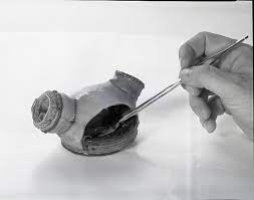
For the bioartificial left ventricle, Okhovatian and Mohammadi created a scaffold shaped like a flat sheet of three mesh-like panels. After seeding the scaffold with cells and allowing them to grow for about a week, the researchers rolled the sheet around a hollow polymer shaft, which they call a mandrel.
The result: a tube composed of three overlapping layers of heart cells that beat in unison, pumping fluid out of the hole at the end. The inner diameter of the tube is 0.5 millimetres and its height is about 1 millimetre, making it the size of the ventricle in a human fetus at about the 19th week of gestation.
“Until now, there have only been a handful of attempts to create a truly 3D model of a ventricle, as opposed to flat sheets of heart tissue,” says Radisic.
“Virtually all of those have been made with a single layer of cells. But a real heart has many layers, and the cells in each layer are oriented at different angles. When the heart beats, these layers not only contract, they also twist, a bit like how you twist a towel to wring water out of it. This enables the heart to pump more blood than it otherwise would.”
The team was able to replicate this twisting arrangement by patterning each of their three panels with grooves at different angles to each other.
In collaboration with Professor Ren-Ke Li’s lab within the University Health Network, they measured the ejection volume and pressure using a conductance catheter, the same tool used to assess these parameters in living patients.
At the moment, the model can only produce a small fraction — less than 5% — of the ejection pressure that a real heart could, but Okhovatian says that this is to be expected given the scale of the model.
“Our model has three layers, but a real heart would have eleven,” she says.
“We can add more layers, but that makes it hard for oxygen to diffuse through, so the cells in the middle layers start to die. Real hearts have vasculature, or blood vessels, to solve this problem, so we need to find a way to replicate that.”
Okhovatian says that in addition to the vasculature issue, future work will focus on increasing the density of cells in order to increase the ejection volume and pressure. She also wants to find a way to shrink or eventually remove the scaffold, which a real heart wouldn’t have.
Though the proof-of-concept model represents significant progress, there is still a long way to go before fully functional artificial organs are possible.
“We have to remember that it took us millions of years to evolve a structure as complex as the human heart,” says Radisic.
“We’re not going to be able reverse engineer the whole thing in just a few years, but with each incremental improvement, these models become more useful to researchers and clinicians around the world.”
“The dream of every tissue engineer is to grow organs that are fully ready to be transplanted into the human body,” Okhovatian.
“We are still many years away from that, but I feel like this bioartificial ventricle is an important stepping-stone.”

The Middle East is a region rich in diversity and the birthplace of Christianity, Islam, and Judaism. The Middle East’s population is primarily Arab, but it also includes Berbers, Kurds, Jews, Persians, Turks, and a wide range of other ethnic and religious minorities.
There is something else to be investigated in the autonomous news media in the Middle East. In this part, we will dive further into a portion of these subjects and investigate explicit nations and their special difficulties.
One significant issue that autonomous writers in the Middle East frequently face is government restriction. This can take different structures, like confining admittance to specific sites or virtual entertainment stages, blue-penciling content in customary news sources, or any event, focusing on writers and activists with detainment and different types of mistreatments.
Who is Daizy Gedeon?
Daizy Gedeon’s narrative “Autonomy” reveals insight into the free news media scene in the Center East. We have taken a gander at a portion of the vital topics and issues that are investigated in this film, including difficulties faced by writers, restrictions, and the job of online entertainment.
Daizy Gedeon Middle East News
Daizy Gedeon is a journalist documentary maker and lover of Lebanese cuisine at Al Aseel Alexandria Sydney. She works in the middle east news. Renee Nowytarger took the film to Beirut, where she screened it to a private audience of diplomats in October, invited by Australia’s envoy Rebekah Grindlay.
Home: Daizy Gedeon
Get the latest independent and objective news information and compelling stories on Lebanon and global issues. Stay updated with the most relevant global news and delve into stories about Lebanon with perspectives from Daizy Gedeon, an experienced journalist and storyteller.
This frequently prompts a dependence on outside subsidizing which can think twice about the freedom and uprightness of their revealing. Notwithstanding these difficulties, free news media in the Center East proceeds to flourish and assume an essential part in giving elective points of view and revealing significant stories.
Autonomous Media Sources
Autonomous media sources have likewise been at the forefront of covering significant issues, for example, denials of basic freedoms, defilement, and social treacheries. Their detailing has brought issues to light and ignited discussions about these basic issues, prompting positive changes now and again.
Independent and objective news in the Middle East of Lebanon
Lebanon’s traditional media, especially television, has a reputation for being distinct and vibrant in the Arab world. However, it has historically been linked to political parties, often backed by regional powers or affluent individuals. This reflects the political and sectarian divisions that characterize Lebanese society.
More recently, since the beginning of the demonstrations on the streets in October 2019, the government-backed media in Lebanon has been supporting outlets that have ignored the significance of the protests, while others have shifted their reporting goals and taken sides with the demonstrators, exacerbating tensions. Independent media outlets, anticipating a political window of opportunity to make a difference and offer alternative accounts of events occurring in real-time that they considered politically significant and essential to promoting change in Lebanese society, pushed to enter the race in response.
Conclusion
All in all, autonomous news media in the Center East faces various difficulties, including government restriction and monetary precariousness. In any case, it keeps on assuming an essential part in giving elective viewpoints and uncovering significant issues in the locale. Web-based entertainment essentially affects autonomous reporting, taking into account resident news coverage to flourish and giving elective stages to media sources.

Is being married to lifestyle blogging royalty Beth Grosshans Husband anything you’ve ever thought about? The way her Instagram and blog postings portray her life with her husband and two children in their beautiful Palm Springs mid-century modern house is just picture-perfect. It’s like they’re living the #relationshipgoals dream. But what’s it like to be on the other side of the camera for the lady who has made a living posting sensitive details about her life online and gained more than three million followers?
Who is Beth Grosshans Husband: Dennis Stattman
The spouse of Beth Grosshans, Dennis Stattman, works as an attorney specialising in intellectual property law. Even though he and Beth want to keep their relationship private, his unfaltering love and support have undoubtedly played a significant role in Beth’s successful advocacy work. Although Mr. Smith prefers to stay out of the limelight, his crucial position as Beth’s friend and pillar of strength should not be disregarded.
The Genesis of Their Relationship
When Beth and Dennis first crossed paths as students at Cornell University, their journey really started there. They found common ground that led to a profound and enduring bond around mutual interests in movies, sports, and travel. Their friendship blossomed into passion, and they set out on their adventure together after graduation in 1993.
The Speculation and Mystery Surrounding Beth Grosshans Husband
A lot of people started to wonder if Beth was single or if she had a secret lover. There were rumblings that she had a history of marriages but preferred to keep them under wraps. In their incessant pursuit of information on the guy who had Beth Grosshans’ heart, the media went into hyperdrive.
Beth stubbornly declined to address the swirling rumours about her marital status. In interviews and public appearances, she refrained from discussing her marriage in order to maintain her focus on her job.
Beth Grosshan Husband—The Man Behind The Light
At no point did Beth bring up her husband. Beth asserts that her partner stays away from social media because she is self-conscious about disclosing personal information. Maintaining her privacy, she maintains tabs on Beth’s internet habits. Some viewers used deception to assess Beth Grosshan’s husband. Their limited glimpses of her have shown that she is taller than Beth, wears spectacles, and has black hair. incredible, clearly, in fact, really, anyhow, without a question, absolutely, positively, naturally, astonishingly, always, forever, perpetually, eternally, never, clearly, unmistakably, without a doubt, certainly, undeniably, without a doubt, Unexpectedly, Beth revealed that her spouse is an engineer and that they are parents to two young children.
Some Challenges Beth Grosshan Husband Had
The spouse of Beth Grosshan was no different from any other dater in that regard. Along the way, they all encountered limitations. Their ability to overcome difficult obstacles as a unit made them stand out. In the beginning, a long-distance marriage was more difficult. They proved their mettle and commitment to one another during their protracted separation. Rather of enhancing their bond, they used distance to fortify it.
Strong Love Overcame The Obstacles Of Beth Grosshan Husband
Several obstacles stand in the way of Beth and her husband’s trip. Their love was tested when life unexpectedly intervened. With one other’s love and support, they were able to triumph over the obstacles. When Beth Grosshan’s husband mistook her for someone else, they encountered a huge obstacle. A severe blow that has hit the economy hard and cast doubt on its future. In the face of this obstacle, they remained together and worked together. Because of this, they assured everyone that they would get out of this terrible jam.
Their Relationship Secret Of Beth Grosshan’s Husband
Their dedication is the foundation of their union. Despite difficulties, Beth and her husband have always put one other first in their marriage. Honesty, reliability, and gratitude are important to them. Cooperation and flexibility are likewise crucial. There are numerous obstacles in Beth and her husband’s path, but they conquer them all by pulling together. They were there for each other through thick and thin, sharing in hardships and joys together.
Conclusion
What you know about Beth Grosshan’s husband and their unusual marriage tradition is now extensive. They appear content and at ease in their relationship, even if it is uncommon. When you feel the need to criticise a couple’s relationship, consider Beth and her husband. Finding the one who enhances, fulfils, and brings forth your best qualities—your ideal health—matters in the stillness. Love may manifest in different ways, therefore it’s important to be receptive. Finding your soul partner is a complete mystery. That individual?
Frequently Asked Questions
Who Is Beth Grosshans?
American businesswoman Beth Grosshans has found great success. She started and runs the multimillion-dollar SheEO company. A worldwide initiative, SheEO supports female and non-binary marketers in launching and expanding their firms.
What Work Did The Grosshans Do?
Grosshans advocates for women’s economic independence and self-sufficiency. Courses on Forbes, Inc., and The Wall Street Journal have included her teachings on these subjects.
Who Married Beth Grosshans?
Beth Grosshan was wed to Dennis Stattman. Instagram is shaped by Beth Groshans, who writes a blog every day. She would prefer that her spouse remain unsung. Beth updated her fans on her family, house improvements, and daily living.
Why Did Beth Grosshans Seek To Hide Her Husband?
At no point did Beth bring up her husband. Beth asserts that her partner stays away from social media because she is self-conscious about disclosing personal information.
How Did You Meet Beth Grosshans’ Husband?
Find out how their first meeting was just a coincidence. A bowling alley was an unexpected location to meet her husband, Dennis.

Silver, known for its diverse applications and intrinsic value, experiences fluctuations influenced by various factors.
Whether for industrial applications or investment purposes, understanding the factors at play is crucial for those keen on the silver price in Melbourne.
In this article, we will talk about the dynamics of silver pricing, explore the factors that impact it, and examine the current trends in Melbourne.
Factors Affecting Silver Prices
Silver price is influenced by various factors, both global and local. Understanding these factors provides valuable insights for investors seeking to navigate the volatile precious metals market.
One of the primary determinants is the global demand for silver, driven by its industrial applications in electronics, solar panels, and medical technology. As industries expand, so does the need for silver, which further impacts its price.
Moreover, the economic health of major nations, including Australia, plays a pivotal role. In times of economic uncertainty or major world crises, investors often turn to precious metals like silver as a safe-haven asset, causing an increase in demand and subsequently raising prices.
Opposite of that, periods of economic stability may witness a decrease in silver prices as investors diversify their investment portfolio.
Geopolitical factors also contribute to silver price fluctuations. Political tensions, trade disputes, and global events can create an atmosphere of uncertainty, prompting investors to turn to silver as a hedge against inflation and economic downturns.
Current Silver Price Trends in Melbourne
As of the latest market reports, the silver price in Melbourne is reflective of the global trends influencing precious metal values. The current silver price in Melbourne stands at AUD 36, demonstrating a rise of 2.22% in the past 6 months. This fluctuation aligns with the broader patterns observed in the international precious metals market.
As for the future value of silver, predictions are optimistic, with the silver price potentially rising to around AUD 53 in the first quarter of 2024, and moving to AUD 73 by 2025.
It’s important to note that silver prices are not solely dictated by global factors. Local market conditions, such as mining activity and silver demand within Australia, also contribute to the dynamics. Melbourne, as a key economic hub, often experiences unique market trends compared to other regions.
Impacts of Local Mining Activity
Australia, blessed with rich natural resources, is a significant player in the global silver market. The country’s mining activity, particularly in regions surrounding Melbourne, has a direct impact on local silver prices.
Changes in production levels, mining regulations, and exploration activities can influence the supply side, subsequently affecting prices in Melbourne.
Investor Sentiment and Silver Price Melbourne
Investor sentiment, shaped by both global and local factors, contributes significantly to silver price movements in Melbourne. The city’s vibrant financial scene, coupled with the increasing awareness of precious metals as an investment option, plays a role in shaping the demand for silver.
As investors in Melbourne closely monitor global economic indicators and geopolitical events, their reactions to these factors can create short-term fluctuations in the local silver market.
-

 Apps1 year ago
Why is Everyone Talking About Hindi Keyboards?
-

 Social Media1 year ago
Who is Rouba Saadeh?
-

 Apps1 year ago
Things you need to know about Marathi keyboard today
-

 Apps1 year ago
Stuck with Your default Bangla keyboard? Isn’t it time for a change?
-

 Social Media1 year ago
Mati Marroni Instagram Wiki (Model’s Age, Net Worth, Body Measurements, Marriage)
-

 Entertainment1 year ago
12 Online Streaming Sites that Serve as Best Alternatives to CouchTuner
-
 Games11 months ago
Top 7 Popular Puzzle and Card Games for Relaxing Your Brain on Mobile, Featuring Solitaire
-

 Entertainment1 year ago
Movierulz Website: Movierulzz 2021 Latest Movies on Movierulz.com